
NEWS
Scattered Inhibitory Clones Carry Forebrain Disorder-Risk Genes
A UCSF and Allen Institute clonal atlas of the newborn mouse forebrain shows inhibitory lineages scatter with myelin precursors and enrich for.

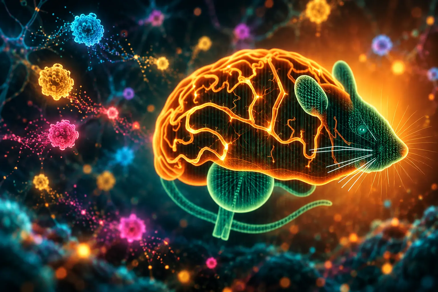
A 30 September 2026 study mapped clonal families across the newborn mouse forebrain and found a split rule for how those families settle. Excitatory neurons stay near astrocytes in the region where they were born. Inhibitory neurons scatter, share ancestry with oligodendrocyte precursor cells, and their far-traveling clones are enriched for genes tied to neurodevelopmental disorders.
Guohua Yuan, a researcher in Tomasz J. Nowakowski’s laboratory at the University of California, San Francisco, led the work with colleagues at the Allen Institute for Brain Science, including Hongkui Zeng. Arturo Alvarez-Buylla, a UCSF developmental neuroscientist whose earlier studies described how radial glia seed adult stem cells, is a coauthor. The paper was received on 8 July 2025 and accepted on 19 August 2026.
Five Labeling Windows, One Newborn Brain
Most lineage maps of the mammalian forebrain have stayed inside the cerebral cortex. Yuan and Nowakowski applied lentiviral barcoding across cortex, hippocampus, striatum, thalamus, and olfactory bulb, then read each cell’s gene activity and its inherited tag in one measurement. They injected the virus from embryonic day 11 through the day of birth and collected tissue at postnatal day 4, when many labeled cells had already taken on mature identities.
The tool is STICR, a barcoding system Nowakowski’s group designed so a unique DNA tag sits in the 3′ UTR of a GFP reporter and can be captured by standard 3′ single-cell RNA sequencing. Libraries deposited at Addgene carry 50 to 70 million unique sequences, enough that two unrelated progenitors are unlikely to draw the same tag. Ryan Delgado, credited in the acknowledgements for the original design, had used the same library to show that individual human cortical progenitors can produce both excitatory and inhibitory neurons.
THE LABELING CALENDAR
- Embryonic day 11: First in utero injections stamp mitotic progenitors along the ventricles.
- Embryonic days 12 to 16: Later embryonic windows catch mid-neurogenesis, including lateral ganglionic eminence output into striatum and olfactory bulb.
- Postnatal day 0: Birth-day labeling marks cells still dividing as the pup is born.
- Postnatal day 4: All brains are collected, dissociated, and sorted for GFP before single-cell sequencing.
- 16 November 2025: Yuan presents the atlas in a Society for Neuroscience talk in San Diego, months before the paper appears.
To put those clones back on a map, the team used MERFISH, a spatial method that reads thousands of genes on intact coronal sections, and registered dissociated profiles onto that anatomy. Quality-control plots cover 93 samples and track cell loss through dissection, sorting, and sequencing. Sampling simulations, 500 runs per condition, asked how many multicellular clones are needed before a lineage’s cell types and fate biases can be trusted.
Excitatory Clones Stay Put With Astrocytes
Glutamatergic neurons, the cells that drive activity in cortex, hippocampus, thalamus, and parts of the olfactory bulb, share clones with astrocytes and remain inside their region of origin. A cortical clone stays cortical. A hippocampal clone stays hippocampal. That pattern extends the old radial-unit picture, in which neurons born from a local radial glial cell stack in a column near their birthplace, and it now includes the astrocyte siblings of those neurons.
Timing still follows the textbook inside cortex. Early labels are enriched for deep-layer projection neurons. Later windows pick up upper-layer neurons and glia. The team also found glutamatergic cells labeled after birth, then checked them with BrdU given at P0 and read at P4 in three animals. Those late excitatory neurons are not a general cortical afterthought. They concentrate in restricted zones, including olfactory bulb populations that can be tagged in the embryo, after birth, or in both windows.
Hippocampal excitatory clones split among dentate gyrus, CA1, and CA3, with only partial barcode sharing across those subfields. Transcriptomic look-alikes from different regions were not, as a rule, clonally coupled. Same-looking excitatory types from two areas usually came from different families.
The Inhibitory Lineage That Will Not Stay Home
GABAergic neurons follow a different bookkeeping. They are clonally related to oligodendrocyte precursor cells, the myelin-making lineage, and they disperse across regional and structural borders. The finding sits on top of 2015 work by Christian Mayer, Corey Harwell, and colleagues showing that clonally related interneurons spread widely. The new atlas places that spread in a whole-forebrain frame and ties it to glial fate.
TWO LINEAGE RULES AT P4
| Lineage | Glial partner | Where sister cells end up |
|---|---|---|
| Glutamatergic neurons | Astrocytes | Kept inside the region of origin |
| GABAergic neurons | Oligodendrocyte precursor cells | Dispersed across forebrain regions |
Marker genes still sort inhibitory clusters toward medial, caudal, or lateral ganglionic eminence programs, but clone sharing does not stop at those embryonic addresses. UpSet plots of GABAergic barcodes show intersections that cross cortex, striatum, and olfactory bulb, and the authors displayed every intersection larger than 200 clones. Cosine distances in gene-activity space were computed for six clone categories, including groups of 4,103 and 4,885 clones, to test whether scattered families are more mixed in identity than stay-at-home ones.
Microglia, endothelial cells, and other non-neural types appear in the dataset, yet they do not obey the same neuron-glia split. Microglia clones are large and wide, consistent with a small set of early myeloid founders, a pattern already described in other barcoding studies of the mouse brain.
How Ventral and Dorsal Striatum Part Ways
The striatum, the main input hub for movement and reward, is where the atlas breaks newest ground. Dorsal and ventral medium spiny neurons, the principal projection cells of the region, are not generated as one program. The paper reports previously unrecognized differences in how those two territories are built, and it links late-born medium spiny neurons to striatal astrocytes and to distinct olfactory bulb neuron pools.
STRIATAL AND OLFACTORY CLONAL GROUPS
- Early striatal core: One GABAergic group is enriched at embryonic day 14 and matches lateral ganglionic eminence neurons that settle in the striatal core.
- Late striatal-OB stream: A second group arises between embryonic days 12 and 16, lasts until P0, and looks transcriptionally like migrating olfactory bulb neurons.
- Early bulb interneurons: Two lateral-eminence-derived olfactory bulb clusters are labeled from embryonic day 11 to 16, peak at 11 to 12, and are gone from P0 collections.
- Late local or V-SVZ group: A second olfactory bulb group is enriched at embryonic day 16 and P0 and likely comes from the ventricular-subventricular zone or from local divisions.
That last point has a timing catch. Olfactory bulb GABAergic neurons collected after P0 labeling shared very few clones with striatum or cortex. Many of the inhibitory cells sitting in the bulb at P4 were born in the embryo or generated nearby. Neurons still migrating through the rostral migratory stream had largely not arrived. The atlas therefore undercounts the postnatal wave that will fill the bulb in the following days, a wave Alvarez-Buylla’s work has long placed in the subventricular zone.
At P0 the team also split postnatal glial and stem-like “B cell” clusters by wall of origin. Cluster 21 (Crym+) mapped as ventral subpallial, cluster 2 (Gsx2+, Dio2+) as dorsal subpallial, and clusters 26, 25, 27, and 1 (Emx1+, Hopx+, Thap2c+) as pallial. Those labels matter for anyone who wants to know which adult stem cell pool a late clone still belongs to.
Disorder Genes Ride the Wandering Clones
The clinical hook is not a new autism gene. It is a spatial pattern. Clones of GABAergic neurons with broad regional dispersion are enriched for genes implicated in neurodevelopmental disorders, including autism-associated genes. The paper points to rare-variant catalogs such as the 2022 analysis by Jack Fu and colleagues, which found 72 autism-associated genes at a false discovery rate of 0.001 in 63,237 people, and to assembloid CRISPR screens of disease genes in human neural cells. Yuan’s atlas does not claim those mutations make cells wander. It shows that the lineages most likely to seed many regions are the ones whose gene programs overlap disease risk.
glutamatergic neurons and astrocytes are clonally related and retained within their region of origin, whereas GABAergic neurons are clonally related to oligodendrocyte precursor cells but disperse extensively across brain regions.
Guohua Yuan, Tomasz J. Nowakowski and colleagues
If a mutation hits a stay-at-home excitatory clone, the damage can stay local. If it hits a dispersing inhibitory clone, sister cells may already be parked in cortex, striatum, and olfactory bulb. That is a developmental way to think about why the same risk gene can touch mood, movement, and sensory circuits at once. It is also a mouse finding. Human cortical progenitors, in Delgado’s STICR xenografts, can produce both excitatory and inhibitory neurons from one parent cell, a mix mouse clones in the related 2022 Bandler study did not show. Anyone using the new atlas as a human template has to keep that species gap in view.
A Map Built for Identity, Place and Time
Viral barcoding captures only a slice of each family, so the authors treated missing sisters as a sampling problem, not a footnote. They built ground-truth clones, then asked how often partial recovery would fake a fate bias, defined as more than 25 percent of cell-type pairs showing no shared clones or a shared-clone fraction below 10 percent of the smaller count. Thalamic dissections included epithalamus and may have picked up a little neighboring tissue, a caveat written into the cluster-level heatmaps.
WHAT THE ATLAS COUNTED
- Multicellular clones: 4,106, 3,348, 1,898, 2,468, and 1,165 clones across the five injection dates, 12,985 in all, after restricting to families with at least two recovered cells.
- Library handling: 93 samples in the cell-loss plots, with GFP-positive cells sorted before every single-cell library.
- Open data: Transcriptomes and barcodes in the Gene Expression Omnibus under GSE341480, deposited 31 July 2026.
- Code and images: MERFISH volumes at the Brain Image Library (dataset ace-let-lax) and custom STICR analysis code on GitHub, with a second repository for the P4 atlas.
An interactive single-cell and MERFISH browser is hosted at the University of California, Santa Cruz. Funding came from the National Institute of Mental Health and the National Institute of Neurological Disorders and Stroke (U01MH130962, R01NS123263, R01MH128364), a California Institute for Regenerative Medicine award of $1,551,394, the Allen Institute, and private foundations. Nowakowski is a New York Stem Cell Foundation Robertson Neuroscience Investigator.
The atlas is a P4 snapshot, not a movie through adulthood. Postnatal olfactory-bulb immigrants are still on the road. Human lineage rules already disagree in one important place. What the resource does give the field is a shared family tree for five forebrain regions, with barcodes, locations, and birth windows attached to the same cells.
Frequently Asked Questions
What is STICR barcoding and how does it tag brain cells?
STICR is a lentiviral library that encodes a CAG-GFP transgene plus an error-correctable barcode in the 3′ UTR, so ordinary oligo(dT) single-cell platforms such as 10x Genomics 3′ chemistry can read lineage and transcriptome together; three indexed pools (Addgene 180483, 186334, and 186335) let labs keep infections from different experiments from mixing.
Which mouse forebrain regions does the clonal atlas cover?
The dissections target cortex, hippocampus, striatum, thalamus, and olfactory bulb, and the thalamic samples also contain epithalamus, with the authors warning that a small amount of adjacent tissue may have been included in those cuts.
Do mouse and human cortical progenitors follow the same excitatory-inhibitory rule?
In this mouse atlas, glutamatergic neurons and GABAergic neurons obey separate clonal partnerships, matching earlier STICR work in which mouse clones did not mix those two classes, whereas Delgado’s 2021 human cortical progenitor study recovered both excitatory and inhibitory neurons from a single parent cell.
Where can researchers download the newborn mouse lineage data?
Single-cell and barcode files live in GEO accession GSE341480, MERFISH images at Brain Image Library record ace-let-lax, the UCSC cell browser hosts the interactive view, and analysis code is split between the NOW-Lab/STICR and guohua-git/p4-brain-lineage GitHub repositories.
Why are so few postnatal olfactory-bulb interneurons in the P4 clones?
Cells labeled at P0 and collected at P4 inside the bulb share almost no barcodes with striatum or cortex, which the authors read as evidence that many P4 bulb interneurons were born earlier or locally, while the postnatal cohort was still in the rostral migratory stream and had not yet arrived.
The browser is live, the barcodes are public, and the split between stay-at-home excitatory families and wandering inhibitory ones is now a number other labs can query. What they do with the disorder-gene overlap is the next experiment, not a result this P4 map can finish.

Final Fantasy Resonance Turns a Dead Gacha Into HD-2D

PHP2Shell Forced Sites Offline While Exploits Stalled

Kindle+ Folds AI Audiobooks Into the Page-Read Fund

Chip Stocks Fall as the Bond Rally Cheapens AI Spend

Comcast’s Record Odyssey Quarter Doubles as the Exit

Microsoft 365 Auth Fault Took Down Exchange and Teams

Xbox Caps Game Pass Cloud Gaming at 15 Hours

OpenClaw 2.0 Ships a Team Workplace With Host-Trust Defaults

OpenAI Unveils GPT-6 Astra With a Critical Cyber Label

Diesel Breaks Its Record as the White House Claims Credit
-
 NEWS2 months ago
Microsoft 365 Auth Fault Took Down Exchange and Teams
-
 GAMING2 months ago
Xbox Caps Game Pass Cloud Gaming at 15 Hours
-
 NEWS2 months ago
OpenAI Unveils GPT-6 Astra With a Critical Cyber Label
-
 NEWS2 months ago
OpenClaw 2.0 Ships a Team Workplace With Host-Trust Defaults
-
 BUSINESS2 months ago
Diesel Breaks Its Record as the White House Claims Credit
-

 LIFESTYLE2 months ago
Squishy Dumpling Toys Recalled After Hiding Illegal Water Beads
-

 BUSINESS2 months ago
ChargePoint Stock Rally Prices Wilmer’s Three-Year Cash Plan
-

 AUTO2 months ago
Tesla Puts Its Wheel-Free Cybercab on Austin Streets